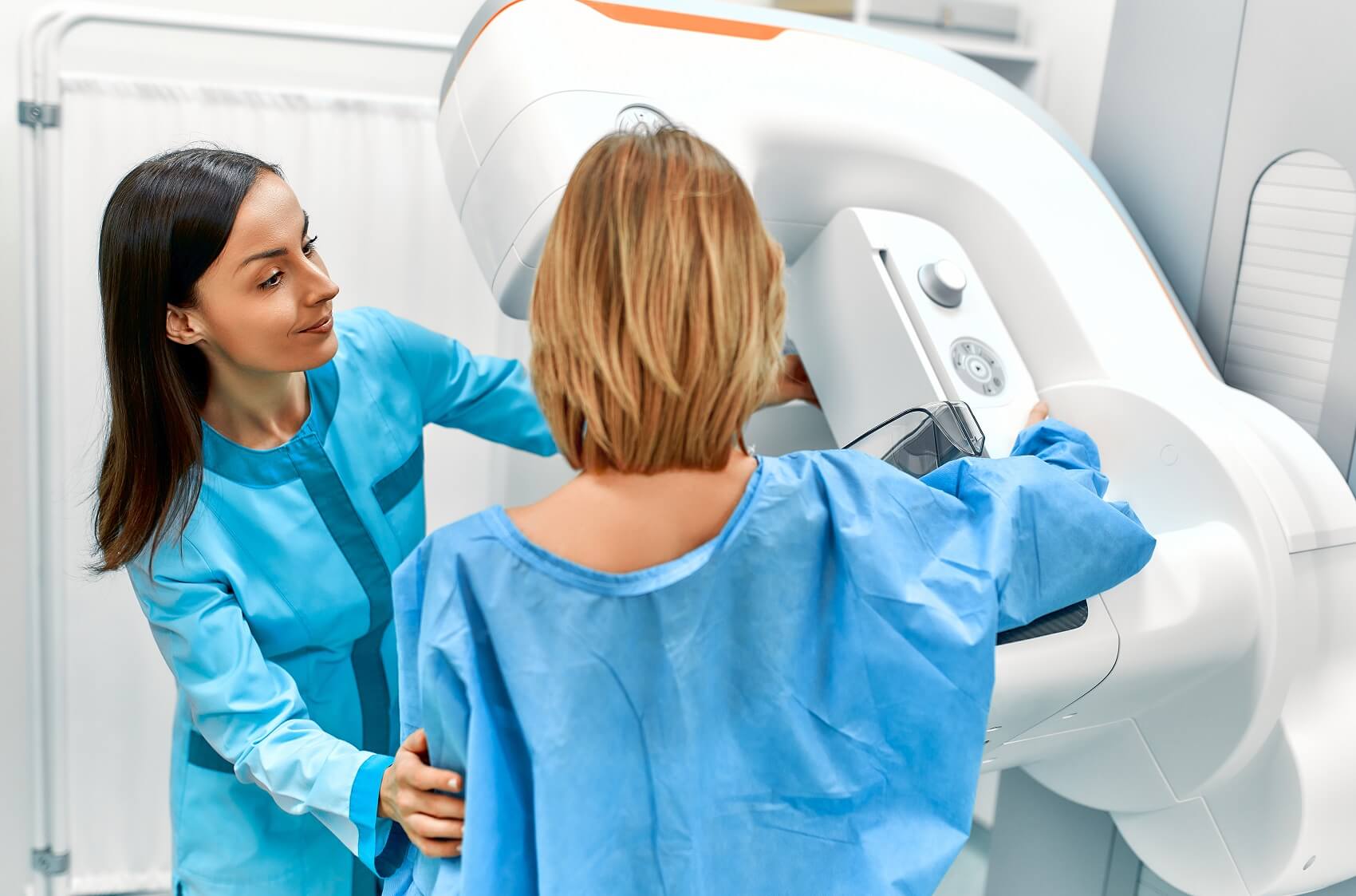
Mammogram
3D mammography, or breast tomosynthesis, is a relatively new breast imaging procedure approved by the U.S. Food and Drug Administration in 2011.
Like traditional mammography, 3D mammography uses X-rays to produce images of breast tissue in order to detect lumps, tumors or other abnormalities. 3D mammography is capable of producing more detailed images of breast tissue.
The 3D mammography procedure resembles that of traditional mammography. The procedure takes place in a private room and is conducted by a radiologic technologist. The woman undergoing 3D mammography is required to remove any clothing above the waist, as well as any jewelry or other objects that might interfere with the imaging process. During the procedure, the woman is positioned before a 3D mammography machine and her breasts are held in place by two compression plates.
The pressure placed on the breasts by the compression plates can cause discomfort but only lasts for a few seconds. When ready, the radiologic technologist will start the 3D mammography machine and a robotic arm will move in an arc over the woman’s breasts as multiple X-ray images are taken. The dose is similar to film mammography and is only slightly higher than in standard 2D digital mammography. The scan itself takes less than two to three seconds per view. The entire procedure takes approximately 10 to 20 minutes.
A radiologist will interpret the results of 3D mammography, looking for signs of calcification or masses in the breast tissue, and report findings according to a standardized classification system known as Breast Imaging Reporting and Data System (BI-RADS). A woman and/or her primary care physician will be notified if her 3D mammography reveals that she has dense breasts or an abnormality in her breast tissue. If an abnormality is detected, a Breastlink Women’s Imaging radiologist will likely recommend additional imaging procedures to help provide women with greater clarity and determine the cause of any true abnormality.
