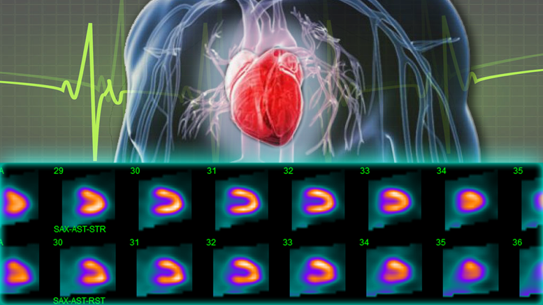
Nuclear Stress Test
What is a nuclear stress test?
Stress tests are diagnostic tests used to evaluate the function of the heart during periods of heavy exercise. During the test, the patient walks on a treadmill (or sometimes uses a stationary bike) at increasing speeds until a target heart rate is reached. Then, imaging tests are performed immediately after the test to evaluate the heart’s performance. A nuclear stress test uses a very small amount of a radioactive dye to highlight the heart, making it easier to see areas of the heart that are not receiving adequate blood flow or are not functioning properly. Once the dye is injected, imaging technology is used to capture still and video images of the heart for evaluation.
When is a nuclear stress test performed?
Nuclear stress tests are performed to diagnose diseases affecting the heart as well to manage treatment of these diseases. Common uses of nuclear stress tests include:
- diagnosing coronary artery disease, which occurs when the arteries supplying blood to the heart become blocked or narrowed, significantly increasing the risk of a heart attack
- evaluating the size of the heart or the thickness of the heart walls
- assessing how well the heart pumps blood
- managing treatment in people diagnosed with heart-related issues
What happens during a nuclear stress test?
Before the stress test begins, the dye will be administered through an IV. Electrodes are attached to the chest to monitor the heart’s activity throughout the stress test. An imaging test is usually performed prior to using the treadmill to obtain baseline data of the heart at rest. The patient will walk on the treadmill until the target heart rate is achieved; then immediately after getting off the treadmill, the heart will be imaged again. Those images can be compared to the pre-test images as well as being evaluated independently. Stress tests are performed on an outpatient basis, and patients can resume their regular activities afterward.
