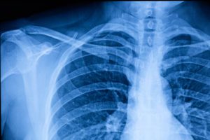
When you think of X-rays, the most common thing to spring to mind is diagnosing broken bones. However, when you look more closely, there is a lot more to X-rays than diagnosing bone fractures.
X-ray is the oldest type of medical imaging technique and still one of the most commonly used methods. X-rays are a type of radiation that can pass through the body to produce images of the structures inside the body. It is a quick, painless procedure that has a variety of uses, including:
- Detecting bone problems such as fractures, arthritis, cancer, and osteoporosis
- Identifying dental problems such as loose teeth, decay, or dental abscesses
- Diagnosing certain types of benign and cancer tumors
- Screening for some types of conditions, such as congestive heart failure, lung cancer, and breast cancer
- Assessing blood vessel health
- Diagnosing lung conditions, such as pneumonia, tuberculosis, or lung cancer
- Identifying digestive tract problems
- Locating an object, such as a swallowed coin or embedded shrapnel
X-rays are very quick and efficient procedures that allow doctors to view and assess results easily, and use the information to help formulate treatment plans.
Facts About X-Rays
- The discovery of X-ray was first reported back in 1895 by German professor, Wilhelm. C. Rontgen. While studying the properties of this type of radiation, he discovered X-ray was able to penetrate ‘screens of notable thickness’.
- Rontgen named it X-ray – the “X” stands for unknown, to indicate that it was an unknown type of radiation, the same as when “X” in mathematics stands for an unknown, or not yet known, value.
- The first use of X-rays under clinical conditions was by Englishman, John Hall-Edwards, in January 1896, when he radiographed a needle that was stuck in the hand of his associate. He was also the first to use X-rays in a surgical operation in February 1896.
- The first medical X-ray made in the United States was carried out in February 1896 by the Frost brothers. It was used to expose the wrist of a patient who was being treated for a fracture. The image of the broken bone was collected on gelatin photographic plates obtained from a local photographer, who was also interested in Röntgen’s work.
- X-ray beams pass through the body and are absorbed in different amounts, depending on the type of material they travel through. X-rays find it more difficult to pass through dense materials, such as bone and metal, as these show up as clear white areas on the image, while softer tissues, such as muscle, appear as shades of gray. Air, such as in the lungs, shows up as black.
- Waves in the electromagnetic spectrum, such as X-rays, can travel through a vacuum at the speed of light.
- In 2009, the X-ray was voted the most important modern discovery by participants in the science museum of London poll. Penicillin came in second.
Types Of X-Rays
X-ray imaging technology has advanced over the years, and nowadays, digital X-ray is a quicker, safer, more cost-effective, and more efficient type of X-ray compared with traditional X-ray imaging. Digital X-ray, unlike conventional film X-ray that uses an intermediate cassette, uses X-ray sensitive plates that directly capture data during the exam and transmit the data to a specialized computer system. Although traditional X-ray is considered safe, digital X-ray emits up to 80% less radiation, making it a much safer alternative. It also provides much clearer, high-quality images compared to traditional X-rays, making it more effective in diagnosing a range of conditions.
There are a variety of X-ray imaging tests available that are used for different purposes. They include:
- Contrast X-rays, which use a contrast agent to help show soft tissues, such as blood vessels and the digestive tract, more clearly.
- CT scans, use multiple X-rays taken from different angles to create cross-sectional, detailed images of tissues and organs inside the body.
- A mammogram, an X-ray picture of the breast, is used to identify the early signs of breast cancer. 3D mammogram combines multiple breast X-rays in various layers to create more detailed three-dimensional images of the breast.
- DEXA (Dual-energy X-ray absorptiometry), uses X-ray beams with different energy levels to measure the density of bones. They are mostly used to evaluate the bone mineral density and to diagnose osteoporosis.
X-ray Diagnostic Services in Bronx, New York
If you are in need of an X-ray or other diagnostic services, the highly qualified team of specialists at Starling Diagnostics can help. We are dedicated to providing the highest quality patient care and offer a wide range of advanced diagnostic radiology services, including digital X-ray, 3T MRI, CT scans, 3D mammograms, and DEXA bone density scans, to support the most comprehensive analysis of musculoskeletal, neurological, and women’s healthcare issues.
To find out more about our radiology services or arrange an appointment, call us at (718) 319-1610. Alternatively, you can use our convenient, secure online request form.
